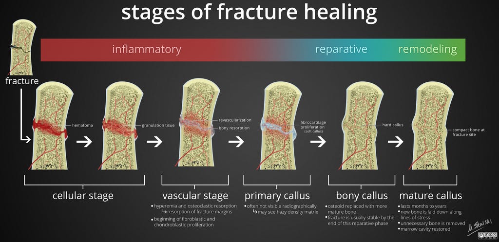
A fracture can be defined as a break in the structural continuity of bone, followed by a loss of integrity (Delforge, 2002). Healing of bone fractures contains within it 3 primary stages: hematoma/inflammation, cellular proliferation, and remodeling (Delforge, 2002). Such stages are comparable to soft tissue healing which include inflammation, fibroplasia, and scar formation (Delforge, 2002).
Understanding the healing process would provide insight into the proximal and distal effects of immobilization, a technique often used during the repair of long bones. Thus, the following will consider the work of Ceroni et al. (2013), and their findings from adolescents recovering from lower limb fractures.

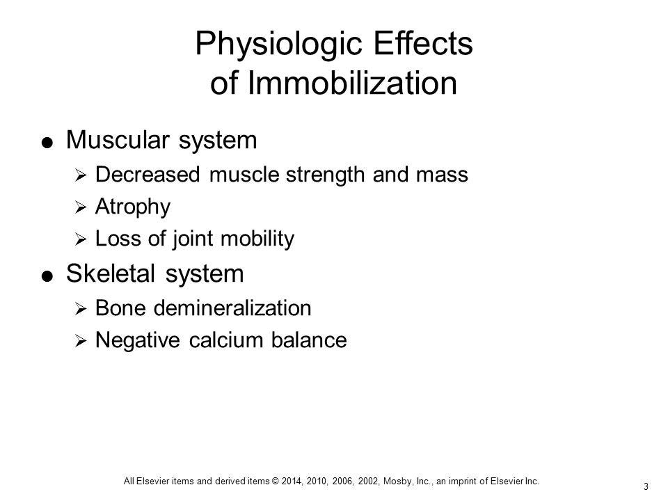
As noted in the aforementioned section, the stages of bone healing are similar to that of soft connective tissue. One particular difference between the aforementioned tissues, however, is that bone regenerates (i.e., restores the original biomechanical properties) while soft connective tissue undergoes scar formation (Delforge, 2002). Immobilization, however necessary, does have side effects; Ceroni et al. (2013) indicated that such an intervention lead to bone loss, atrophy, and functional limitations.
Other key facts included losses not only in bone mineral density (BMD) at the fracture site, but also in more distal regions of the same long bone (the researchers examined fractured femoral bones). A prominent finding of Ceroni et al. (2013) included BMD loss as high as 50% in adolescents during the first 6-12 months following fracture. At 18 months, however, BMD (measured by dual x-ray absorptiometry) was similar to that of the contralateral limb, and control subjects.

Although research has indicated BMD is fully restored in adolescents after 18 months (a seemingly long period), the aforementioned recovery time is considerably faster than adults recovering from the same fracture (Ceroni et al., 2013). Although the intent of Ceroni et al. (2013) was to demonstrate the side effects of immobilization, and the rapid rate of recovery in adolescents compared to adults, one could still submit it takes a long time for a bone to fully recover, despite age group.
Such knowledge has encouraged this author to consider the time and place of exercise and load-bearing activity; all forms of exercise progressions post-injury (i.e., sets, repetitions, weight, exercise complexity) should be implemented in a slow and pragmatic fashion. In this way, restoring BMD, hypertrophy, and activities of daily living are expedited in a manner that compliments the stages of healing, while being efficient, and safe.
References
Ceroni, D., Martin, X.E., Delhumeau, C., Farpour-Lambert, N.J., De Coulon, G., Dubois-Ferrière, V., & Rizzoili, R. (2013). Recovery of decreased bone mineral mass after lower-limb fractures in adolescents. Journal of Bone and Joint Surgery, 95-A(11), 1037-1043.
Delforge, G. (2002). Musculoskeletal trauma; Implications for sports injury management. Champaign, IL: Human Kinetics.
-Michael McIsaac
