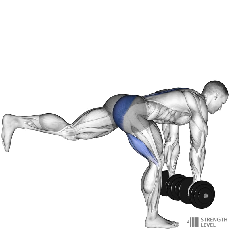
The back and knee joints are regions often susceptible to injury. The hip musculature, when working dysfunctionally, is associated with back pain (McGill, 2010). The hips are also implicated with poorly functioning and painful knees (Bolga, Malone, Umberger, & Uhl, 2008). The deadlift, when performed correctly, is an exercise that has been shown to recruit the gluteus muscles optimally and restore proper function (Bolga et al., 2008). Due to the mechanics required of the deadlift, the low back and knees require little to no motion. This allows stimulation of the hip musculature while circumventing larger motions, and potential pain, at the hips, low back and knees. In an effort to more deeply appreciate this exercise, I would like to explore the deadlift in different phases of the pattern. I will also provide the relevant anatomy including muscle actions, joints and planes of motion involved. Finally, I will show how to develop and progress the deadlift into a single leg version safely and pragmatically. Below is a movement analysis of the single leg deadlift with video links:
Single Leg Deadlift (Front View)
Single Leg Deadlift (Side View)
Activity: Single Leg Deadlift
Phase 1: Moving Downward
| Joint
(closed chain leg) |
Start Position | Movement | Plane | Axis | Muscle/Action |
| Ankle(talocural joint) | neutral | static | n/a | n/a | Isometric Contraction
Plantar Flexors:
Prime Movers: gastrocnemius, soleus
Synergists: fibularis longus, fibularis brevis, plantaris, tibialis posterior, flexor digitorum longus, flexor hallusic longus
Dorsi Flexors:
Prime Movers: anterior tibialis
Synergists: extensor hallucis longus, extensor digitorum longus, fibularis tertius |
| Knee (patellofemoral joint) | extended | flexion | sagittal | mediolateral | Co-contraction
Knee Flexors:
Concentric Contraction:
Prime Movers: semitendonosis, semimembranosis, biceps femoris
Synergists: sartorius, popliteus, gracilis
Knee Extensors:
Eccentric Contraction:
Prime Movers: rectus femoris, vastus lateralis, vastus intermedius, vastus medialis
Synergists: tensor fascia latae |
| Hip (acetabulofemoral joint) | extended | flexion | sagittal | mediolateral | Co-contraction
Hip Flexors:
Concentric Contraction: Prime Mover:
Synergists: iliacus, rectus femoris, sartorius, tensor fascia latae, pectineus, gluteus minimus.
Hip Extensors:
Eccentric Contraction:
Prime Movers: gluteus maximus
Synergists: semimembranosus, semitendonosis, biceps femoris (long head), gluteus medius (posterior). |
| Low Back (lumbar vertebrae) | neutral | static | n/a | n/a | Isometric Contraction
Lumbar Flexors: (rectus abdominis, external oblique, internal oblique)
Lumbar Extensors:
semispinalis, multifidus, and rotatores |
Activity: Single Leg Deadlift
Phase 2: Moving Upward
| Joint
(closed chain leg) |
Start Position | Movement | Plane | Axis | Muscle/Action |
| Ankle (talocural joint) | neutral | static | n/a | n/a | Isometric Contraction
Plantar Flexors:
Prime Movers: gastrocnemius, soleus
Synergists: fibularis longus, fibularis brevis, plantaris, tibialis posterior, flexor digitorum longus, flexor hallusic longus
Dorsi Flexors:
Prime Movers: anterior tibialis
Synergists: extensor hallucis longus, extensor digitorum longus, fibularis tertius |
| Knee (patellofemoral joint) | flexed | extension | sagittal | mediolateral | Co-contraction
Knee Flexors:
Eccentric Contraction:
Prime Movers: semitendonosis, semimembranosis, biceps femoris
Synergists: sartorius, popliteus, gracilis
Knee Extensors:
Concentric Contraction:
Prime Movers: rectus femoris, vastus lateralis, vastus intermedius, vastus medialis
Synergists: tensor fascia latae |
| Hip (acetabulofemoral joint) | flexed | extension | sagittal | mediolateral | Co-contraction
Hip Flexors:
Eccentric Contraction:
Prime Mover:
Synergists: iliacus, rectus femoris, sartorius, tensor fascia latae, pectineus, gluteus minimus.
Hip Extensors:
Concentric Contraction:
Prime Movers: gluteus maximus
Synergists: semimembranosus, semitendonosis, biceps femoris (long head), gluteus medius (posterior). |
| Low Back (lumbar vertebrae) | neutral | static | n/a | n/a | Isometric Contraction
Lumbar Flexors:
rectus abdominis, external oblique, internal oblique
Lumbar Extensors:
semispinalis, multifidus, rotatores |
APPLICATION OF THE SINGLE LEG DEADLIFT
Most activities that individuals experience occur predominantly on one leg such as walking, climbing stairs, and changing direction (McCurdy, O’Kelley, Kutz, Langford, Ernest, & Torres, 2010). When training any movement pattern, the more muscle activity involved, the more force and stability that can be produced. McCurdy et al. (2010) discovered that there was higher EMG activity in the gluteus medius and hamstring musculature in a single leg stance when compared to a bilateral stance. This has value because the gluteus medius helps level the pelvis and help the gluteus maximus control femoral internal rotation, a common movement dysfunction that arises from poorly stabilized hips (Bliven, 2014).
Another incentive to target single leg exercises arises from a group of muscles that help control the knee and hips; the lateral subsystem. This system is composed of the same-side adductor magnus, gluteus medius and contralateral quadratus lumborum (Clark, Lucett, & Kirkendall, 2010). These muscles help control both the hip and knee in the frontal plane while in a single leg stance. If this system is working in a dysfunctional fashion, femoral internal rotation and adduction of the same hip can occur. This equates to a poorly stabilized patellofemoral joint and a lumbar spine that moves excessively in lateral flexion (Clark, Lucett, & Kirkendall, 2010).
The SLD is a movement that can access, train, and strengthen the musculature and subsystems responsible for knee and low back stability. Moreover, it teaches individuals how to lift objects without placing their backs and knees in sub-optimal postures. However, the SLD demands focus, coordination and significant motor control. Complex exercise involves controlling the joints of the body in a coordinated and harmonious fashion. Magill (2011) refers to this as the degrees of freedom problem. Often, novices will become “stiff” and lock out several joints when they are exposed to novel and complex exercises, leading to undesirable and aberrant motions. Magill (2011) explains this as an attempt to control the new movement, though in an inefficient manner.
In an effort to control the degrees of freedom and develop the SLD, I implement the concept of constraints from motor learning theory. Clark (1995) theorized that the environment we are surrounded by shapes movements, analogous to a glass giving shape to water. To assist clients in their training, I developed a repository of videos for their reference. In the following clips, I use constraints (i.e., broom handle along the back and a bench across the knees) to help control the degrees of freedom, develop and “shape” the SLD motion. This process systematically removes the constraints until the person performs the SLD free of assistance
Progressions (click links below for video):
2 Leg Deadlift + Knee and Spinal Constraint + No Load
2 Leg Deadlift + Spinal Constraint + No Load
2 Leg Deadlift + No Constraints + No Load
2 Leg Deadlift + Landing Constraint + Bilateral Load
2 Leg Deadlift + No Constraints + Bilateral Load
2 Leg Deadlift + Landing Constraint + Unilateral Load
Single Leg Deadlift + Contralateral Load + Landing Constraint
Terminal Exercise (click links below for video):
Single Leg Deadlift + Contralateral Load + No Landing Constraint (Side View)
Single Leg Deadlift + Contralateral Load + No Landing Constraints (Front View)
ASSOCIATED RESTRICTIONS
As mentioned, the gluteus maximus and medius are often weak. Another trend seen in modern populations is a condition known as lower crossed syndrome (Page, Frank & Lardner, 2014).Essentially, a crossedpattern of inhibited and facilitated muscles exists between the anterior and posterior regions of the hips: the psoas group of the anterior hip as well as the spinal erectors of the posterior low back are facilitated. Conversely, the gluteus maximus and gluteus medius of the posterior hip and abdominals of the anterior trunk are weak or inhibited. These conditions can worsen control and stability of the low back and knees, as well as performance of the single leg deadlift. If these problems exist, they should be focused upon before developing the single leg deadlift.
Low back and patellofemoral pain can be managed, even mitigated, by restoring proper and well-controlled movement. Understanding the anatomy, pathomechanics and progressions of exercise is paramount. Only then, can we affect change in a manner that is both safe and effective.
References
Bliven, K. (2014). Functional anatomy of the knee. Part 2: Anatomy [Slideshow Presentation]. Retrieved February 16, 2014, fromhttp://assets.atsu.edu/BB/ASHS/HM/HM502/HipThightP2.mp4
Bolga, L. A., Malone, T. R., Umberger, B. R., & Uhl, T. L. (2008). Hip strength and hip and knee kinematics during a stair descent in females with and without patellofemoral pain syndrome. Journal of orthopedic and sports physical therapy. 38 (1), 12-18.
Clark, J. E. (1995). On Becoming Skillful: Patterns and constraints. Research Quarterly for ExerciseSport, 66 (3), 173-183.
Clark, M., Lucett, S., & Kirkendall, D. T. (2010). NASM’s essentials of sports performance training. Baltimore, MD: Lippincott Williams and Wilkins.
Magill, R. A. (2011). Motor learning and control: Concepts and applications (9th ed.). New York: McGraw-Hill.
McCurdy, K., O’Kelley, E., Kutz, M., Langford, G., Ernest, J., & Torres, M. (2010). Comparison of lower extremity EMGbetween the 2-legsquat and modifiedsingle-legsquat in female athletes. Journal of Sport Rehabilitation. 19 (1), 57-70.
McGill, S. (2010). The painful lumbar spine.IDEA Fitness Journal, 7 (1),32-37.
Page, P., Frank, C., & Lardner, R. (2014). The Janda approach to chronic pain syndromes: Preserving the teachings of Dr. Vladimir Janda. Retrieved January 25, 2014 from http://www.jandaapproach.com/the-janda-approach/jandas-syndromes/
-Michael McIsaac
