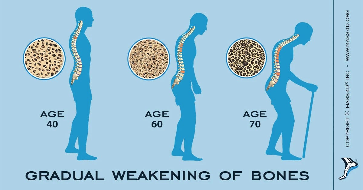
Osteoporosis is a condition characterized by elevated bone turnover and compromised bone strength, which increases an individual’s risk of fracture.1 Furthermore, 44 million Americans are estimated to have low bone mass which, in 2001, had a direct national expenditure of 17 billion dollars.1(1015) Ultimately, early monitoring and tracking of bone mineral density (BMD), and other biomarkers, are critical steps in prevention and management of osteoporosis. As such, the following will consider such metrics, and nutritional interventions, to support optimal bone development and maintenance.

In this author’s previous posts, diagnosis of osteoporosis/BMD can be determined via Duel Energy X-ray absorptiometry scan, commonly known as a DEXA. BMD norms are determined by comparing an individual’s result against a healthy population of the same sex followed by assigning a T-score (measures number of standard deviations from the mean).2Osteoporosis is defined as a T-score at or below -2.5.2(295) Although DEXA is useful, said diagnostic technique is limiting as it does not indicate nutritional/biochemical drivers behind osteoporosis. Thus, close analysis of other biomarkers can help elucidate aberrations in bone density, as well as providing solutions to treating the same.
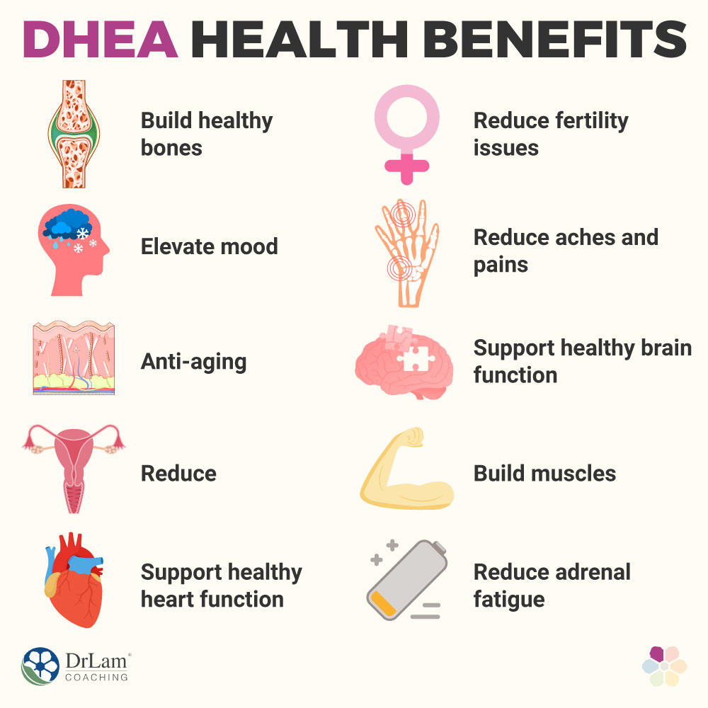
Dehydroepiandrosterone (DHEA) is a precursor to testosterone/estrogen and is involved in tissue development, growth, immune system strength, and cardiovascular function.3 Furthermore and most relevantly, DHEA is associated with muscle strength and bone mineral density, especially amongst females.3(558),4 Thus, if such a marker is not within range (i.e., 3-10 ng/ml) and is specifically low, steps should be taken to regulate and support a return to normal ranges. Interestingly, high levels of stress can negatively affect, and lower, DHEA levels.3(560) Such stress from anxiety/poor sleep/excessive exercise can cause the adrenal glands (where DHEA is made) to produce excessive levels of cortisol. Thus, lowering said stresses could help return DHEA levels to normal ranges.
If DHEA remains low despite managing stresses and having normal cortisol levels (a measure of adrenal response and stress levels), supplementation of DHEA may be indicated. Lord et al3(558) stated that 50 mg/day of DHEA for one year improved hip BMD in older males and spine BMD in older females. Furthermore, when 50 mg/day of DHEA was provided to post-menopausal females, said hormone increased estrone/estradiol/androstenedione/testosterone/DHEA sulfate.3(558) Finally, Lord et al3(561) cited a study indicating adults in their 70s who took 50 mg/day of DHEA for 6 months had improved fat-free mass, lowered fat mass, and improved BMD. Thus, DHEA could serve as an effective hormone replacement therapy.
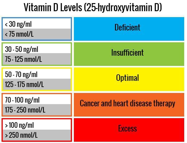
Vitamin D3 (VD3) levels, measured in the blood as 25-hydroxyvitamin D (25OHD), is another key marker related to BMD maintenance. VD3 is a micronutrient that has been associated with significantly reducing all-cause mortality, and has been implicated in many diseases of modern civilization.5 Furthermore, VD3 interacts with multiple organs, to include BMD, and more than 200 genes implying its broad reach and influence upon human physiology.5(6) If 25OHD levels are below range (i.e., <40 ng/ml), supplementation might be indicated if individuals cannot get adequate sun exposure.5(13) Optimal ranges, according to Cannell et al5(13) are considered to be between 40-70 ng/ml. Thus, slowly increasing VD3 and rechecking 25OHD levels are indicated until optimal ranges are ascertained.

Another key metric is undercarboxylated osteocalcin (UCOC); a functional marker of vitamin K (VK) deficiency. Such a measure is relevant since osteocalcin, and UCOC, are proteins produced by osteoblasts (cells that build bone) which are involved in bone density maintenance.7 When UCOC levels are high, such a marker becomes produced from the breakdown and degradation of bone matrix which is then released into the bloodstream.7(446) VK facilitates bone deposition as it carboxylates and binds calcium to bone.3(447) Thus, low VK can translate into higher levels of UCOC, which in turn, increases bone degradation. Therefore, supplementation of VK-rich foods such as leafy green vegetables (i.e., collard greens, spinach, broccoli) and/or supplementation to return UCOC levels to normal ranges.8
In conclusion, osteoporosis is a condition characterized by elevated bone turnover and compromised bone strength, which increases an individual’s risk of fracture. Furthermore, 44 million Americans are estimated to have low bone mass which, in 2001, had a direct national expenditure of 17 billion dollars. Ultimately, early detection (i.e., DEXA) and monitoring of nutrient status (i.e., UCOC, VD, VK, DHEA) are critical biomarkers to consider when supporting management of osteoporosis. When combined with a larger protocol (physical activity, resistance training), such an integrated approach is likely to help slow BMD loss, reduce risk of fractures, and improve quality of life.
References
1. Srivastava AK, Vilet EL, Lewiecki M, et al. Clinical use of serum and urine biomarkers in the management of osteoporosis. Curr Med Res Opin. 2005;21(7):1015-1026. doi:10.1185/030079905X49635.
2. Lee RD, Nieman DC. Nutritional Assessment. 6th ed. New York, NY: McGraw-Hill. 2013.
3. Lord RS, Bralley, JA. Laboratory Evaluations for Integrative and Functional Medicine. 2nded. Duluth, GA: Genova Diagnostics; 2012.
4. Hong SH, Kim JH, Lee JH, et al. Dehydroepiandrosterone sulfate and free testosterone but not estradiol are related to muscle strength and bone microarchitecture in older adults. Calcif Tissue Int. 2019;105(3):285-293. doi:10.1007/s00223-019-00566-5
5. Cannell JJ, Hollis BW. Use of vitamin D in clinical practice. Altern Med Rev. 2008;13(1):6-20. http://archive.foundationalmedicinereview.com/publications/13/1/6.pdf. Accessed March 17, 2020.
6. Rosen CJ. Clinical practice.Vitamin D insufficiency.N Engl J Med. 2011;364(3):248-254. doi: 10.1056/NEJMcp1009570.
7. Zanetta LCB, Boguszewski CL, Borba VZC, et al. Association between undercarboxylated osteocalcin, bone mineral density, and metabolic parameters in post-menopausal women. Arch Endocrinol Metab. 2018;62(4):446-451. doi:10.20945/2359-3997000000061.
8. Gropper SS, Smith JL, Carr, TP. Advanced Nutrition and Human Metabolism. 7th ed. Boston, MA: Cengage Learning; 2018.
-Michael McIsaac
