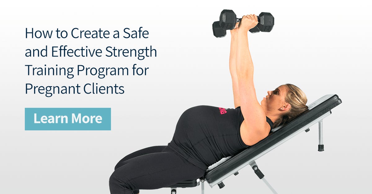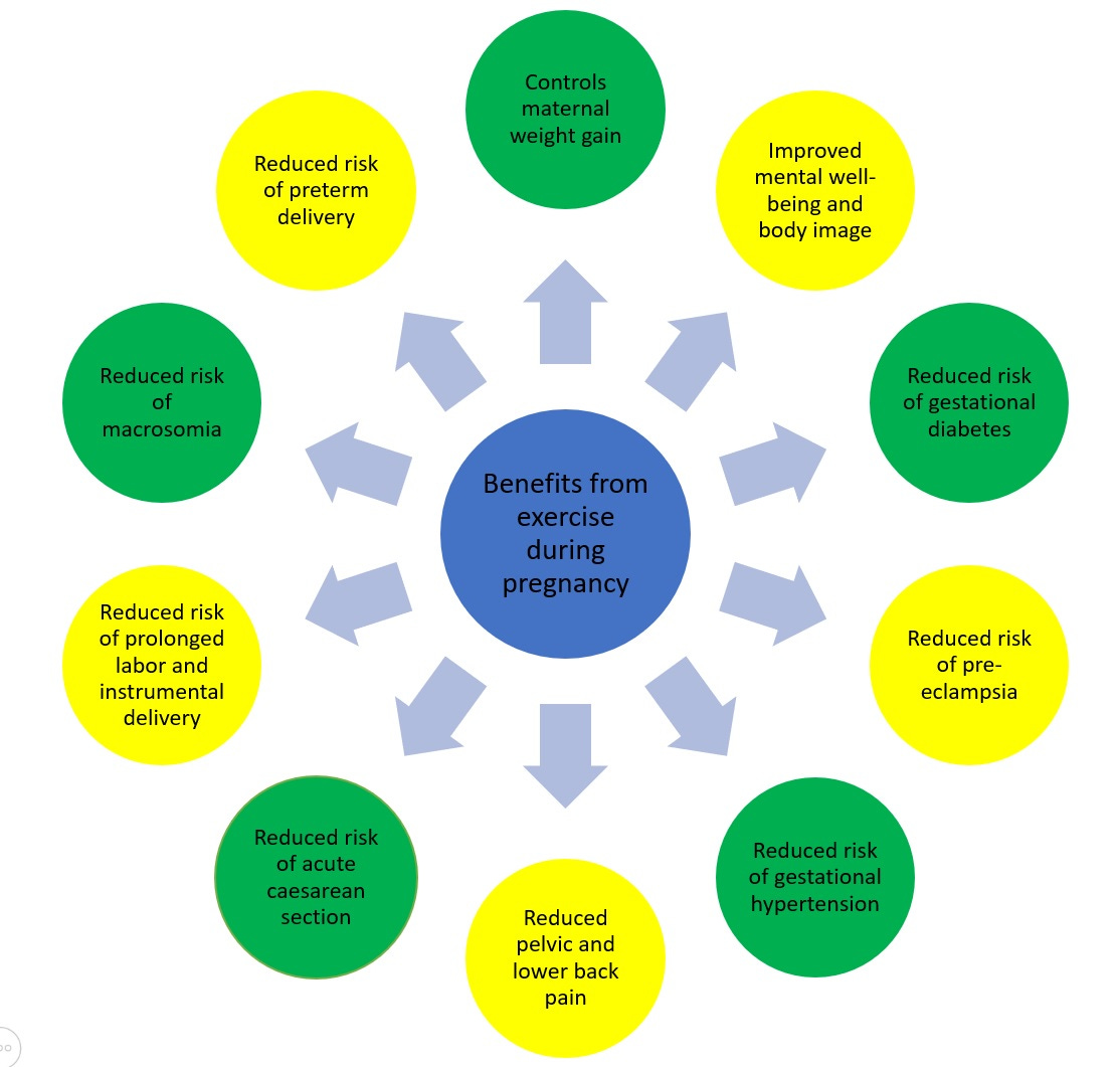
Evidence-based, periodized exercise programs have the capacity to improve several biomarkers such as cardiovascular adaptations, higher bone mineral density, improved blood profiles, and muscular strength (Piper, Jacobs, Haiduke, Waller, & McMillan, 2012). Considered universally favorable adaptations for all people, what remains unique however, is how such improvements are sought for each individual, and special populations. The following sections will review the unique physiological and biomechanical changes of females who are pregnant, as a means of appreciating the demands that exercise places on the mother. Finally, the author will cover contraindicated, and indicated, core exercises based on evidence and best practice.

Piper et al. (2012) suggested that, in addition to the benefits of exercise in the aforementioned section, physical activity also enabled easier labors, shorter labor times, as well as enhanced recovery after birth. Such knowledge creates incentive for pregnant individuals to engage in physical activity. However, like medicine, there are different types of exercise, different doses, and frequencies that elicit specific outcomes; the exercises must to be appropriately matched to the needs and circumstances of the client.

Pregnancy poses unique physiological and biomechanical changes in the female body. As the mother moves from the first to third trimester, abdominal distension induces deeper lordosis, as well as shifting the center of mass forwards. Such changes compromise the mother’s balance and spinal stability (Piper et al., 2012). Other unique alterations include a weakened venous return from the fetus pressing against the inferior vena cava (Piper et al., 2012).
Temperature regulation is also of importance; the baby’s core temperature is generally 1°C higher than the mother. Such an occurrence is of utmost relevance as Piper et al. (2012) noted that an increase of temperature beyond 1.5°C of the mother could elevate the temperature of the fetus to a level which could induce congenital malformations. Considering the changes, which occur in a pregnant female’s body, the following will explore exercises that enhance core stability, while acknowledging the physiological and biomechanical uniqueness of the mother.

Piper et al. (2012) suggested that core stabilization is a crucial component of a mother’s exercise program. Despite exercise choice, several tenets will be upheld during training; the client has to breath during all exercise, which helps guarantee avoidance of the valsalva maneuver. Such an approach will help keep blood pressure from rising too high, as well as maintaining normal venous return.
The environment must be normal room temperature or a little lower, and the client must rest between sets. The aforementioned approaches attempts to keep the mother’s temperature from rising too high, thusly keeping the fetus’ temperature from rising above 1.5°C. All core exercises should be regressed to reduce torque on the anterior core, initially. Since the anterior core is distended, myosin and actin filaments are stretched, which may place them in a weakened position (Baechle & Earle, 2000).

Piper et al. (2012) provided several core exercises whereby there is motion at the lumbar spine (i.e., twists, crunches, side bends). This author chooses to avoid exercises that place tension and loads against the lumbar region, as McGill (2007) provided evidence that repetitive motions in the sagittal, frontal, and transverse planes can cause tissue failure in the discs of the low-back.
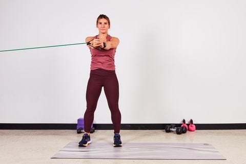
Thus, all exercises chosen by the author are isometric in nature, resisting motions in the frontal, sagittal, and transverse planes. Exercises could include bird-dogs, elevated front planks, and elevated side planks (i.e., transverse plane, sagittal plane, frontal plane, respectively). Level of intensity is determined by incorporating the rate of perceived exertion. Piper et al. (2012) recommended that mothers stay within an intensity, which falls between 12-14, indicating a “somewhat hard” effort that also falls between 50%-80% of maximum heart rate range.
Evidence supports the inclusion of physical activity for females who are pregnant. However, vigilance must be implemented when developing programs and training clients. Each exercise, set, rep, and progression of a periodized regime should be based on evidence and best practice, with sound reasoning behind each aforementioned constituent. Such an approach provides an opportunity to improve not only the health of the mother; it also protects the welfare and growth of the developing fetus.
References
Baechle, T.R., & Earle, R.W. (2000). Essentials of strength and conditioning (2nd Ed.): National Strength and Conditioning Association. Champaign, IL: Human Kinetics.
McGill, S. (2007). Low back disorders: Evidence-based prevention and rehabilitation (2nd ed.). Windsor, ON: Human Kinetics.
Piper, T., Jacobs, E., Haiduke, M., Waller, M., & McMillan, C. (2012). Core training exercise selection during pregnancy. Strength and Conditioning Journal, 34(1), 55-58.
-Michael McIsaac
