In my last post, I spoke about the need to incorporate single leg training to enhance the stability of the hip and knee musculature. In the following paragraphs, I would like to explore how distal joints and tissues may influence knee stability, as well as their implications on patellofemoral pain syndrome.

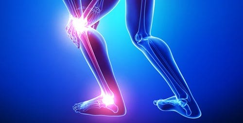
Most injuries from extracurricular activities occur within the lower extremities. 42% of these are directly related to the knee (Macrum, Bell, Boling, Lewek, & Padua, 2012). One of the mechanisms of injury is thought to be excessive motion of the patellofemoral joint in the frontal plane (Macrum et al., 2012). Thus, understanding these motions, and more importantly, their etiologies, is the first step in learning how to treat these aberrant movement patterns.

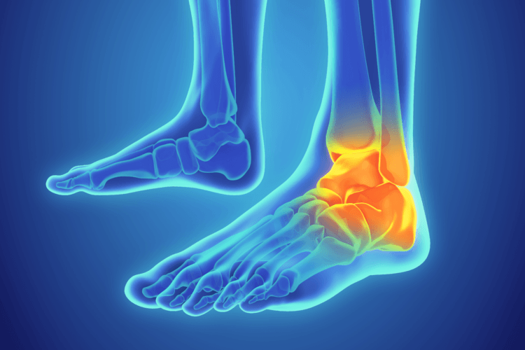
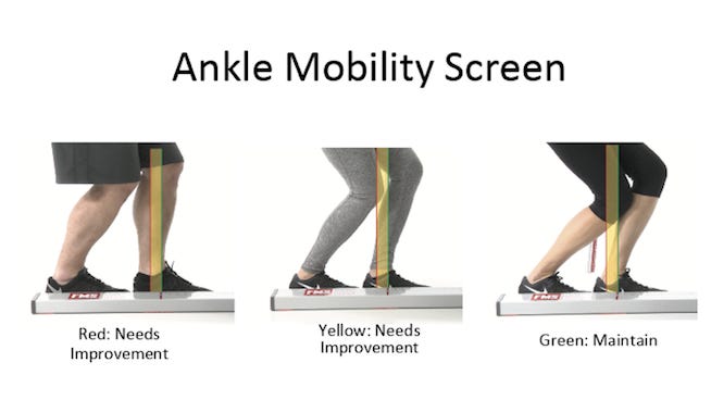
Macrum et al. (2012) analyzed the ankle joint to see how restricted dorsiflexion could affect leg kinematics and muscle activation patterns. Of interest is the association between patellofemoral pain and ankles; most people who have knee pain lack dorsiflexion (Macrum et al., 2012). Additionally, Macrum et al. (2012) noted that reduced dorsi flexion caused the foot to pronate, the tibia to internally rotate, causing femoral internal rotation and valgus collapse at the knee.

The study had 15 men and 15 women perform squats in induced dorsiflexion with a foot wedge. EMG and motion analysis was performed. They discovered that reduced dorsiflexion caused a loss in knee flexion and increased valgus collapse, as well as a loss in quadriceps activation. This causes weakness and increased difficulty in performing the squat. Reduced dorsiflexion, reduced knee flexion, and increased valgus collapse as a group have also been implicated as postures related to patellofemoral pain syndrome (Macrum et al., 2012).
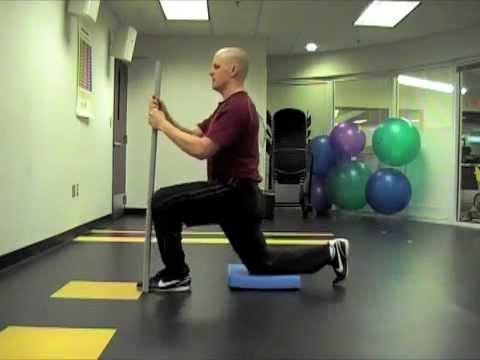
In conclusion, the ankle dorsiflexion pattern is important, and we have seen that the loss of this motion is associated with knee instability. These conclusions should encourage us to think about how we can restore mobility of the ankle. More importantly, how might we intervene so that dorsiflexion is not lost at all?
References
Macrum, E., Bell, D. R., Boling, M., Lewek, M., & Padua, D. (2012). Effect of limitingankle-dorsiflexion range of motion on lower extremity kinematics and muscle-activation patterns during a squat.Journal of Sport Rehabilitation. 21(2), 144-150.
Effect of limiting ankle-dorsiflexion range of motion on lower extremity kinematics and muscle-activation patterns during a squat
-Michael McIsaac
