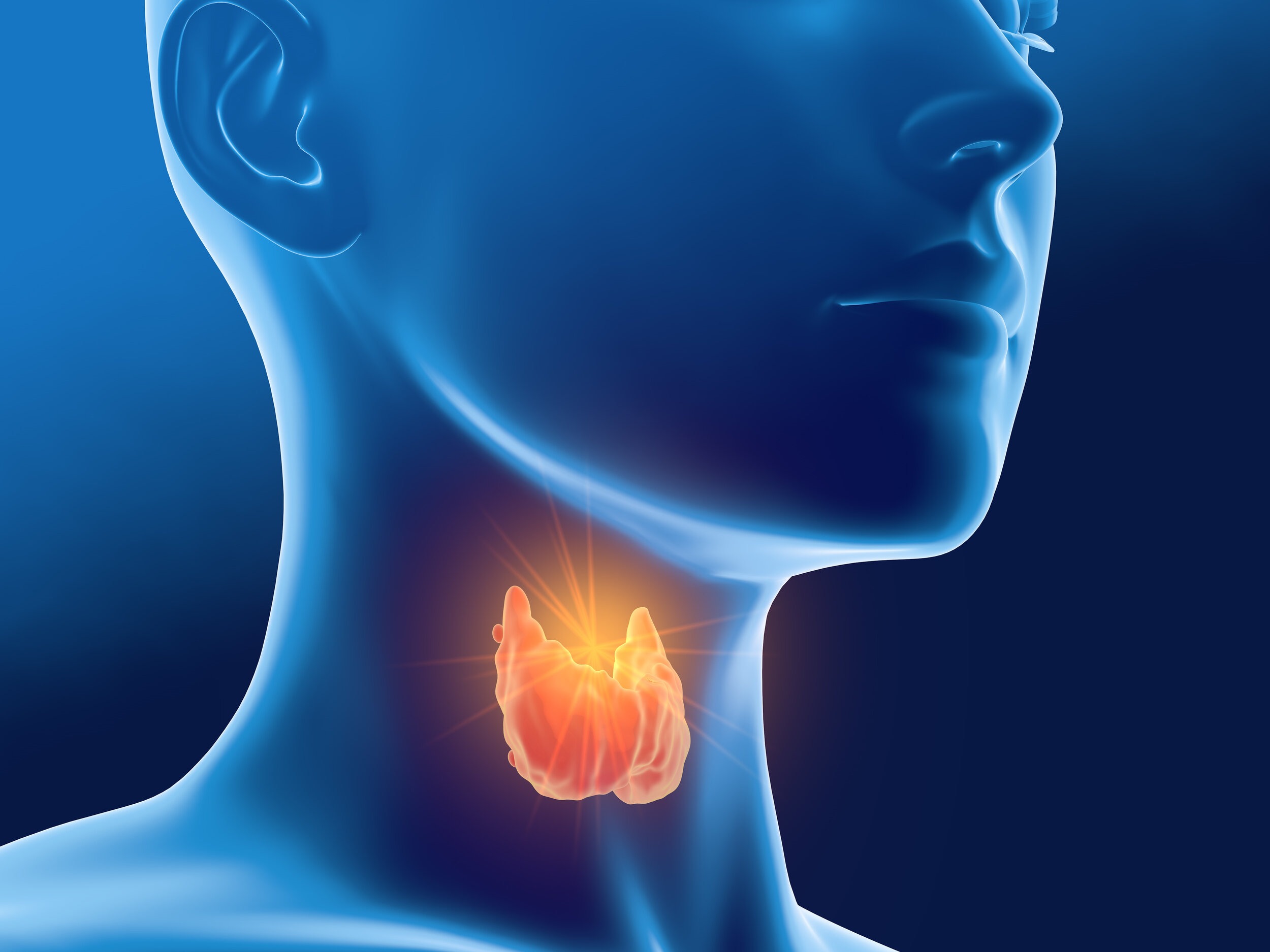
Metabolic processes, in addition to normal growth and development, are heavily influenced by the endocrine system; a group of organs, which release hormones directly into the bloodstream (Reisner & Reisner, 2017). Such endocrine glands include the pituitary, adrenal cortex, medulla, pancreas, kidneys, parathyroid, and thyroid. Aberrations in the performance and function of said glands can negatively affect homeostasis, ranging from barely detectable signs and symptoms to extreme dysfunction (Reisner & Reisner, 2017). As a means of appreciating the role endocrine glands play in maintaining health and longevity, the following will briefly consider the thyroid gland, its hormones, and roles within the body.
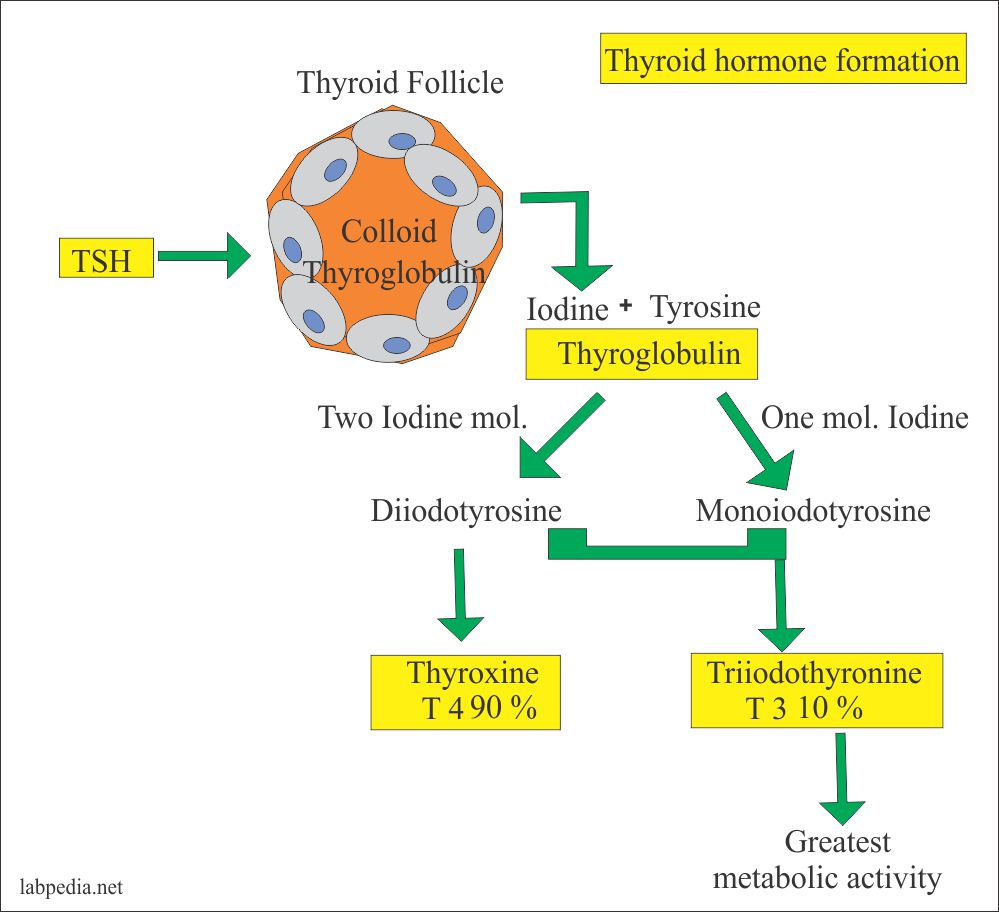
The thyroid is a gland that sits along the upper part of the trachea consisting of two lateral lobes. Within the gland are follicles which harbor a binding protein (thyroglobulin) that attaches to thyroid hormone; the substance responsible to regulating energy levels and metabolic processes throughout the body (Reisner & Reisner, 2017). Thyroid hormone can be broken down into two main types: thyroxine (T4) and triiodothyronine (T3). T4 is formed by the binding tyrosine (an amino acid) to iodine forming monoiodotyrosine. Another iodine molecule is incorporated forming diiodotyrosine which eventually binds to another diiodotyrosine molecule forming tetraiodothyronine (T4). When a diiodotyrosine binds with a monoiodotyrosine, triiodothyronine is formed (T3) (Pagana & Pagana, 2014). Such a conversion mainly occurs in peripheral tissues such as the liver (T4 is converted to T3) which is facilitated by a selenium-dependent enzyme (Thyroid Regulation, 2018). Having considered the two main types of thyroid hormone, the following will consider two subtypes; free T4 and free T3.
T4 and T3 are general terms that describe total quantities of both hormones in the body. However, said hormones are made inactive until required by the tissues in the body. As a means of controlling interactions T4 and T3 with cell membranes, both hormones are bound to carrier proteins, known as thyroid binding globulins, that inhibit their activity (Reisner & Reisner, 2017; Thyroid Regulation, 2018). Since the body does require some T4 and T3, small amounts flow throughout the body unbound to carrier proteins, allowing said hormones to interact and influence metabolism and cellular activity. Such unbound T4 and T3 molecules are known as free T4 and free T3, respectively (Thyroid Regulation, 2018). When individuals are in homeostasis, the endocrine glands function in a balanced and coordinated fashion. However, any disruption in said systems can cause aberrations in T4 and T3 levels. The following will explore the same.
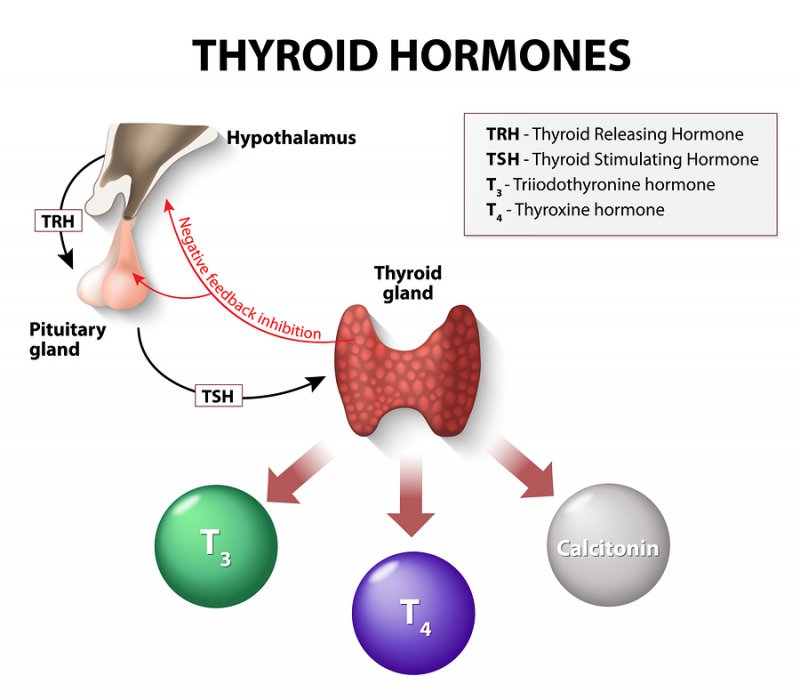
Thyroid hormone levels are closely regulated by the hypothalamus and the pituitary gland. T4 and T3 concentrations are managed by a feedback loop; the hypothalamus releases thyrotropin releasing hormone (TRH) which stimulates the pituitary gland to release thyroid stimulating hormone (TSH) (Thyroid Regulation, 2018). TSH stimulates the thyroid gland to release T4 and T3. When T4 and T3 levels rise, the hypothalamus monitors said levels in the blood and begins down regulating the release of TRH, thereby reducing the release of TSH from the pituitary, ultimately reducing the release of T4 and T3 from the thyroid gland (Thyroid Regulation, 2018). Under normal conditions, such a feedback loop tightly controls T4 and T3 levels. However, such a process can become disrupted by another form of hormone known as reverse T3 (RT3).
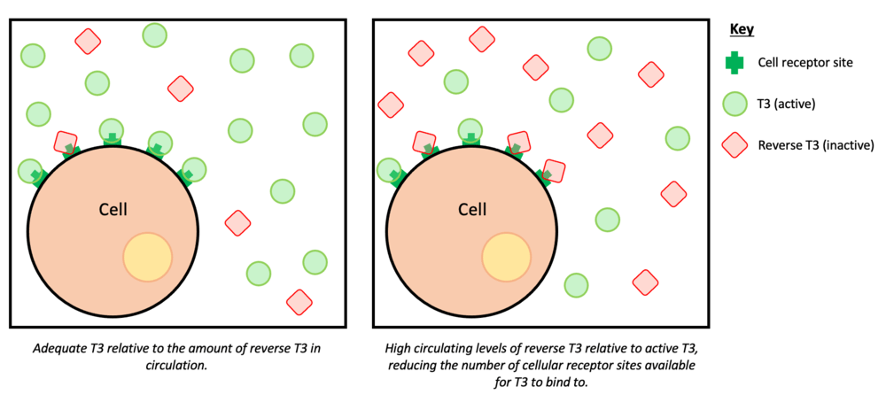
In some individuals, total T4 and T3 levels can look normal (4500 ng/dL-11,700 ng/dL, and 80-200 ng/dL, respectively) in the presence of hypothyroidism (Test ID: T4, 2018; Test ID: T4, 2018). Such an anomaly can be explained by a molecule known as RT3; RT3 differs from T3 in the positions of the iodine atoms attached to the aromatic rings. The majority of RT3 found in the circulation is formed by peripheral deiodination of T4 (Test ID: RT3, 2018). RT3 binds to cells of the body that T3 normally interacts with. However, RT3 is considered inactive, and cannot participate in delivering oxygen and energy to tissues requiring the same. In effect, RT3 competes for placement (against T3) on cell membrane receptors. High levels of RT3 can lower metabolism and overall energy in individuals. Furthermore, stress and extreme exercise can elevate RT3 and downregulate TSH, thereby lowering T3 (Thyroid Regulation, 2018).
In conclusion, the endocrine system works in a harmonious and synchronized fashion to maintain health and homeostasis. However, thyroid function can become compromised under conditions of extreme stress (voluntary or involuntary) creating dysregulation of hormone production and utilization, as seen in the production and role of RT3.
References
Pagana, K. D., & Pagana, T. J. (2014). Manual of diagnostic and laboratory tests (5thed.). St. Louis, MO: Mosby.
Reisner, E. G., & Reisner, H. M. (2017). An introduction to human disease: Pathology and pathophysiology correlations (10thed.). Burlington, MA: Jones & Bartlett Learning.
Test ID: RT3 (2018). Mayo Clinic. Retrieved from https://www.mayomedicallaboratories.com/test-catalog/Clinical+and+Interpretive/9405
Test ID: T3 (2018). Mayo Clinic. Retrieved from https://www.mayomedicallaboratories.com/test-catalog/Clinical+and+Interpretive/8613
Test ID: T4 (2018). Mayo Clinic. Retrieved from https://www.mayomedicallaboratories.com/test-catalog/Clinical+and+Interpretive/36108
Thyroid Regulation (2018). Life Extension. Retrieved from http://www.lifeextension.com/Protocols/Metabolic-Health/Thyroid-Regulation/Page-01
-Michael McIsaac
