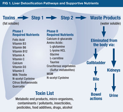
The liver has the capacity to manage toxic exposures via a layered process; phase 1 and phase 2. Such a process begins by the production of important enzymes (i.e., cytochrome p450) made within liver cells that help expedite the reduction/oxidation/hydrolysis reactions (i.e., phase 1) (Grant, 1991; Kim & Lee, 2006). Phase 2 is characterized by conjugation reactions. As an aggregate, phase 1 and 2 processes convert and prepare toxic substances for excretion. However, conditions such as sepsis can severely hamper the production of cytochrome p450 (CP450) and overall detoxification processes via overproduction of reactive oxygen species (ROS). As such, it is imperative CP450 production and activity is increased. The following will consider how vitamin C (VC) and vitamin E (VE) would help in such a process.
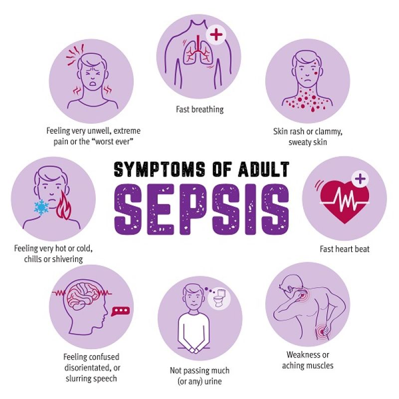
Kim and Lee (2006) indicated that there is sufficient evidence to suggest sepsis is a condition that induces oxidative stress. The researchers also indicated that antioxidants can inhibit the cellular communication and unfavorable gene activation that ROS can initiate, with particularly influential affects from VC and VE (Kim & Lee, 2006). For example, VC was stated as a powerful electron donor that could “feed” superoxide and hydroxyl radicals (i.e., ROS) (Kim & Lee, 2006).VE is a lipid-soluble micronutrient that can protect cells from the damaging effects of free oxygen radicals (i.e., VE donates its hydrogen atom) and facilitates expression of CP450 (Kim & Lee, 2006).
Interestingly, individuals suffering from sepsis have unusually low/depleted levels of VC and VE. To explore the efficacy of said antioxidants, the researchers used rat models whereby sepsis was induced via cecal ligation and puncture. Kim and Lee (2006) had rats randomly assigned to the following groups:
(a) saline-treated control (n = 3)
(b) soybean oil-treated control (n = 3)
(c) VC-treated control (n = 3)
(d) VE-treated control (n = 3)
(e) saline-treated cecal ligation and puncture (n = 6)
(f) soybean oil-treated cecal ligation and puncture (n = 6)
(g) VC-treated cecal ligation and puncture (n = 10)
(h) VE-treated cecal ligation and puncture (n = 10)
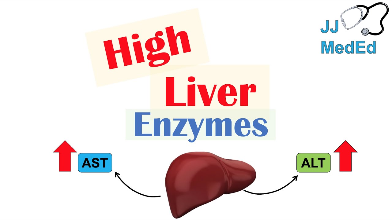
Markers of ROS activity was measured before and after each intervention. Such markers included alanine aminotransferase (ALT), which is a measure of liver damage and is a screen for liver disease, while aspartate aminotransferase (AST) is another enzyme that indicates liver damage and liver disease (Kim & Lee, 2006). Lipid peroxide levels were also considered, as they are a measure of lipid cell membrane damage from ROS (Kim & Lee, 2006). Glutathione (GSH) (an antioxidant) and glutathione disulfide levels (GD) were monitored, since GD is the oxidized form of the antioxidant, indicating its use against ROS (Sadhu, Xie, Zhang, Perumal, & Guan, 2016). Finally, CP450 reductase (helps transfer electrons from NADPH to CP450) was also measured.
Results indicated that all cecal ligation groups experienced heightened levels of ALT and AST levels. However, VC and VE groups experienced lowered levels of the same biomarkers, indicating that such antioxidants are helpful in improving liver function (Kim & Lee, 2006). Furthermore, lipid peroxide levels were lower in the VC and VE groups compared to the other cecal ligation groups, indicating the influence of said antioxidants in controlling oxidative stress (Kim & Lee, 2006). GSH levels remained higher in VC groups compared to all other groups, suggesting that VC spares GSH and behaves as a first line of defense (Kim & Lee, 2006). Finally, CP450 was low in cecal ligation groups devoid of VC and VE, while VC and VE experimental groups experienced a prevention of CP450 loss and maintained NADPH-CP450 reductase levels (Kim & Lee, 2006).

In conclusion, VC and VE share a close relationship with other antioxidants (i.e., GSH) and helps attenuate cell and organ damage. Most importantly, said antioxidants reduce the reach and damage of ROS throughout the body by donating their electrons. Such evidence should help encourage the inclusion of said micronutrients in the diet.
References
Grant, D. M. (1991). Detoxification pathways in the liver. Journal of Inherited Metabolic Disease, 14(4), 421-430.
Kim, J. Y. & Lee, S. M. (2006). Vitamins C and E protect hepatic cytochrome P450 dysfunction induced by polymicrobial sepsis. European Journal of Pharmacology, 534(1), 202-209.
Sadhu, S. S., Xie, J., Zhang, H., Perumal, O., & Guan, X. (2016). Glutathione disulfide liposomes- A research tool for the study of glutathione disulfide associated functions and dysfunctions. Biochemistry and Biophysics Reports, 7, 225-229.
-Michael McIsaac
